NOTIFICATIONS

Welcome

Monday, March 13, 2017

ACID FAST STAINING

                                      Acid Fast Stain (Ziehl- Neelsen Method)

Aim:

            To perform acid fast staining to identify Mycobacterium tuberculosis and Mycobacterium leprae.

Introduction :

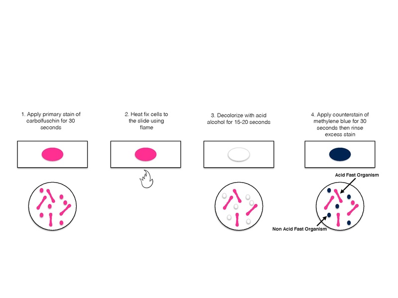
            The Ziehl Neelsen Method is used for staining Mycobacterium sp. in clinical specimens. The thick outer waxy covering (mycolic acid) of the Mycobacterium cell walls act as a barrier  and does not allow all the stains to enter into the cell. In order to visualize these cells higher concentrations of the staining solution is needed and once this stain enters the cell, it is too difficult to remove the stain using a decolorizer. When the clinical specimen is stained with basic dyes such as carbol fuchsin (primary stain) with the continuous application of heat, softens the waxy lipid outer covering of the cell wall and the stain readily enters the cell and stains the cell cytoplasm. When decolorizing agents such as acid-alcohol is added over the primary stain, some bacterial cells cannot be easily decolorized and such bacterial cells are called as acid fast bacteria. The bacteria with high concentration of lipid are easily decolorized by the decolorizing agent and are said to be non-acid fast bacteria. Finally, the addition of the counter stain, Methylene blue, dyes the colorless non acid fast cells as blue thus differentiating them from the pink acid fast bacteria which are unaffected by the Methylene blue.

 Materials Required:

1.      A clean grease free slide.
2.      A bacterial cell suspension.
3.      Staining agent- carbol fucshin.
4.      Boiling water bath.
5.      Decolourising agent – Acid alcohol
6.      Counter stain – 1% Methylene blue


Procedure :
  1. Smear of organisms were prepared on clean glass slide
  2. The slide was allowed to air dry and heat fixed..
  3. The slide was flooded with carbol fucshin stain and placed on a boiling water bath for steaming for about 3-5 minutes.
  4. During steaming the stain is repeatedly added on the slide to avoid drying of smear.
  5. Further the slide was delcolourised with acid alcohol until the stain disappear in washing.
  6. After decolourisation the slide was given a water wash treatment.
  7. Further the smear was flooded with the counter stain that is 1% Methylene blue for about one minute.
  8. The slide was then washed with water, air dried and observed under oil immersion objective.

Result: 

On microscopic observation, the acid fast bacterium appeared as pink coloured cells, non-acid fast cells appeared blue.

Discussion:


            Acid fast stain is due to relative solubility of carbol fuchsin and impermeability of call wall. Fuchsin is more soluble on carbolic acid than in water and carbolic acid soluble more easily in lipids than in acid alcohol. Therefore carbol fuchsin has higher  affinity for lipids than acid alcohol and will remain within the cell wall when washed with decolouriser.


Sunday, March 12, 2017

Screening of PHB (Poly- β- hydroxybutyrate) Producing Bacteria


Aim:

          To Screen the PHB producing Bactria.

Introduction:

          Bacterial Cytoplasm contains a large number of inclusions called cytoplasmic inclusions for example volutin granules, metachromatic granules, phosphate granules,  Poly- β- hydroxybutyrate granules etc. The PHB is active microbial polyester and has been observed in many bacteria including rhizobia. It is an internal reserve food due to which bacterial cells survive to extended period of starvation in soil.

Materials Required:

Bacterial culture (Alcaligenes sp., Rhizopbia and Bacillus Sp.,)
0.3 % Sudan Black B solution (in ethylene glycol)
Safranin
Microscopic slide
Cover slip
Microscope

Procedure:

        1.    Prepared thin bacterial  smear on a slide surface, and thoroughly air dried

2.    Stained the smear with sudan black b solution for 5-15 min.

3.    Drained and dry out the slide and washed the slide with distilled water.

4.    Counter stained the slide with safranin for 10 -20 seconds.

5.    Then rinsed the slide in distilled water and blot dried with tissue paper.

6.    Observed the slide under microscope.  
1. 

Result:
         
          Blue droplets of Poly- β- hydroxybutyrate were observed, while cytoplasm appeared pink in oil immersion microscopy.

ESTIMATION OF CHLOROPHYLL PIGMENTS


Aim:

To estimate the chlorophyll pigments from plant leaves.

Introduction:

The chlorophylls are the essential components for photosynthesis, and occur in chloroplasts as green pigments in all photosynthetic organisms. They bound loosely to proteins but are readily extracted in organic solvents such as acetone or ether etc. Chemically each chlorophyll molecule contains a porphyrin nucleus with a chelated magnesium atom at the centre and a long chain hydrocarbon (phytal) side chain attached through a carboxylic acid group. There are at least five types of chlorophylls in plants. Chlorophyll a and b  occur in higher plants, ferns and mosses. Chlorophylls c, d and e are only found in algae and in certain bacteria.

          Chlorophyll is extracted in 80% acetone and the absorption at 663 nm and 645 nm are read in a spectrophotometer. Using the absorption coefficients, the amount of chlorophyll is calculated.

Material required:

Plant leaves, Mortar and pestle, 80% acetone, centrifuge, standard flask

Procedure:
1              1.   1 g of finely cut and well mixed representative sample of leaf was                                 weighed and took into a clean mortar.

2.    The leaves  were grinded to a fine pulp with the addition of 20 ml 80%    acetone.

3.    The pulp was centrifuged (5,000 rpm for 5 min) and the supernatant was transferred to the 100 ml standard flask.

4.    The residues were mixed and grinded with 80 % of acetone, centrifuged and supernatant was transferred to the same flask.

5.    The procedure was repeated until the residue became  colorless. The mortar and pestle washed thoroughly with 80 % acetone and the clear washing were collected in the standard flask.

6.    The volume of  extract was mad to 100 ml with , 80% acetone

7.    The absorbance of the solution was read at 645,663 and 652 nm against the solvent (, 80% acetone) blank.


Calculation:  (Write Left Side using pencil) 

Calculate the amount of chlorophyll present in the extract mg chlorophyll per g tissue using the following equations.

                mg   chlorophyll a/ g tissue =  12.7 (A663)  - 20.69  (A645
               mg   chlorophyll  b/g tissue =  22.9  (A645)  - 4.68  (A663
               mg   of total chlorophyll /g tissue =  20.2 (A645)  - 8.02  (A663


Result :

          mg   chlorophyll a/ g tissue = 
          mg   chlorophyll  b/g tissue = 

mg   of total chlorophyll /g tissue =  

Friday, March 10, 2017

Fungal Staining by Lactophenol Cotton Blue method (LPCB)

 Aim

To stain fungal cells by using Lactophenol cotton blue.

Principle 

The lactophenol cotton blue (LPCB) wet mount preparation is the most widely used method of staining and observing fungi and is simple to prepare. The preparation has three components:
1.      Phenol:   kills any live organisms;
2.      Lactic acid : It  preserves fungal structures, and
3.      Cotton blue : It stains the chitin in the fungal cell walls.

Lactophenol Cotton Blue Solution is a mounting medium and staining agent used in the preparation of slides for microscopic examination of fungi. Fungal elements are stained intensely blue.

Materials Required

1.      Microscope
2.      Glass Slide
3.      Cover Slip
4.      70 % alcohol
5.      Lactophenol cotton blue (LPCB)
6.      Inoculation needle
7.      Fungal culture

Procedure

  1. Placed a drop of 70% ethanol on a clean microscopic glass slide
  2. The Immersed the specimen in the drop of alcohol
  3. Added  one or two drops of the LPCB before the alcohol dries out
  4. Hold  the coverslip between the index finger and thumb, touch one edge of the drop of mountant with a coverslip edge and lower gently avoiding air bubbles
  5. Made the initial examination using low power objective then Switched to higher power (40X) objective for more detailed examination of spores and other structures.

Observation

After staining fungus cell observed  different parts of a cell like conidia, hypae as well as conidiophore etc.

Result


On basis of observation the type fungal organism identified as Aspergillus sp.,


Alagae, Fungi, Parasite

Entamoeba histolytica 

Entamoeba histolytica is an anaerobic  parasitic  amoebozoa, part of the genus Entamoeba.  Predominantly infecting humans and other primates causing amoebiasis. Inside humans Entamoeba histolytica lives and multiplies as a trophozoite. Trophozoites are oblong and about 15–20 µm in length. In order to infect other humans they encyst and exit the body. The life cycle of Entamoeba histolytica does not require any intermediate host. Mature cysts (spherical, 12–15 µm in diameter) are passed in the feces of an infected human. Another human can get infected by ingesting them in fecally contaminated water, food or hands. 


Aspergillus sp.,
Aspergillus is a genus consisting of a few hundred mold species found in various climates worldwide. spergillum is an asexual spore-forming structure common to all Aspergillus species; around one-third of species are also known to have a sexual stage. Aspergillus is defined as a group of conidial fungi—that is, fungi in an asexual state

Human and animal disease
Aspergillus causes  lung disease, called Aspergillosis, Aspergillosis is, in particular, frequent among horticultural workers who inhale peat dust, which can be rich in Aspergillus spores. It has been found in the mummies of ancient Egyptian tombs and can be inhaled when they are disturbed

Industrial uses

Aspergillus  is cultured for the industrial production of many substances. Various strains of A. niger are used in the industrial preparation of citric acid (E330) and gluconic acid (E574) and have been assessed as acceptable for daily intake by the World Health Organisation. A. niger fermentation is "generally recognized as safe" (GRAS) by the United States Food and Drug Administration under the Federal Food, Drug, and Cosmetic Act.
 Many useful enzymes are produced using industrial fermentation of A. niger. For example, A. niger glucoamylase is used in the production of high-fructose corn syrup, and pectinases are used in cider and wine clarification.
 Some Aspergillus species are
A.niger,  A.flavus , Aspergillus fumigates, A. terreus etc.,


Penicillium Sp.,

Penicillium is a genus of ascomycetous fungi of major importance in the natural environment as well as food and drug production. Some members of the genus produce penicillin, a molecule that is used as an antibiotic, which kills or stops the growth of certain kinds of bacteria inside the body. Other species are used in cheesemaking.
The thallus (mycelium) typically consists of a highly branched network of multinucleate, septate, usually colorless hyphae. Many-branched conidiophores sprout on the mycelia, bearing individually constricted conidiospores. The conidiospores are the main dispersal route of the fungi, and often are green in color.
Economic Value : 
Several species of the genus Penicillium play a central role in the production of cheese and of various meat products. To be specific, Penicillium  molds are found in Blue cheese. Penicillium camemberti  and  Penicillium roqueforti  are the molds on Camembert, Brie, Roquefort, and many other cheeses.  Penicillium nalgiovense  is used to improve the taste of sausages and hams, and to prevent colonization by other molds and bacteria.[17]
In addition to their importance in the food industry, species of Penicillium and Aspergillus serve in the production of a number of biotechnologically produced enzymes and other macromolecules, such as gluconic, citric, and tartaric acids, as well as several pectinases, lipase, amylases, cellulases, and proteases. Some Penicillium species have shown potential for use in bioremediation because of their ability to break down a variety of xenobiotic compounds.
The genus includes a wide variety of species molds that are the source molds of major antibiotics. Penicillin, a drug produced by P. chrysogenum (formerly P. notatum), was accidentally discovered by Alexander Fleming in 1929, and found to inhibit the growth of Gram-positive bacteria (see beta-lactams).


Rhizopus Sp., 


Rhizopus is a genus of common saprophytic fungi on plants and specialized parasites on animals. They are found on a wide variety of organic substrates, including "mature fruits and vegetables",[2] jellies, syrups, leather, bread, peanuts, and tobacco. 
Rhizopus species grow as filamentous, branching hyphae that generally lack cross-walls (i.e., they are coenocytic). They reproduce by forming asexual and sexual spores. In asexual reproduction, sporangiospores are produced inside a spherical structure, the sporangium. Sporangia are supported by a large apophysate columella atop a long stalk, the sporangiophore. Sporangiophores arise among distinctive, root-like rhizoids. In sexual reproduction, a dark zygospore is produced at the point where two compatible mycelia fuse. Upon germination, a zygospore produces colonies that are genetically different from either parent.
·         R. microsporus var. oligosporus is used to make tempeh, a fermented food derived from soybeans.
·         R. oryzae is used in the production of alcoholic beverages in parts of Asia and Africa.
·         Rhizopus stolonifer (black bread mold) causes fruit rot on strawberry, tomato, and sweet potato and used in commercial production of fumaric acid and cortisone.


Chlamydomonas 
Chlamydomonas is a genus of green algae consisting of unicellular flagellates, found in stagnant water and on damp soil, in freshwater, seawater. It is generally found in habitat rich in ammonium salt. Chlamydomonas possesses red eye spots for photosensitivity and reproduces by both asexual and sexual means.
Chlamydomonas's asexual reproduction goes through Zoospores, Palmella stage, Aplanospores and Hypnospores; sexual reproduction through isogamy, anisogamy or oogamy.

Nutrition

Most species are obligate phototrophs but C. reinhardtii and C. dysosmos are facultative heterotrophs that can grow in the dark in the presence of acetate as a carbon source.

Morphology

·         Motile unicellular algae.
·         Generally oval in shape.
·         Cell wall is made up of glycoprotein and non cellulosic polysaccharides instead of cellulose.